3D CT Guided Dental Implant Placement
3D CT Imaging for Guided Dental Implant Placement
Dental implants typically require extensive planning by the surgeon and your general dentist. Dr. Kachele performs both the surgery and the Prosthetic restorations in his office and has been doing this for over 25 years. Is most cases, using a combination of the clinical examination and careful review of your traditional 2-D dental images, i.e. Panoramic X-Ray or Periapical X-Rays of the surrounding jaw and teeth, we are able to gather the majority of the needed information. However in many circumstances, these 2-D images may not be sufficient. This is where 3-D imaging is revolutionizing the treatment of dental implants.
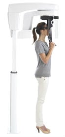
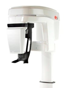
We utilize the use of a combination 2-D / 3-D machine: The Carestream 8100 3D allows us to only obtain the 3-D imaging when needed, reducing your exposure to unnecessary RADs or Radiation.
The 3-D images are then used to determine the safest location for the placement of the dental implant. By having this information prior to surgery we are now able to plug this information into the latest innovation in implant treatment planning software. Using this software we are able to fabricate a surgical guide that will allow us to place the implant with proper surgical planning, down to 0.5mm! This helps to avoid any surprises during the procedure.
This is what we mean by “CT-guided Implant Placement”. The 3D imaging actually allows the procedure to be “performed” in advance of your surgery. A 3-D CT is obtained in our office, then using Anatomage we are able to place the implant or implants within the 3D mock-up of your dentition in the safest and most ideal positions. These implant positions can be shared in advance with your general dentist so that treatment is ideal.

For a single implant placement, this procedure can take less than one hour. This is where planning on our end, helps to save you time. Typically there are no incisions or sutures, leading to less post-operative swelling and discomfort. Most patients only take pain medication for the first few days, although you may be on short-course of antibiotics afterwards.
This state-of-the-art technology can be used for in patients needing 1 implant or 10 implants, in a full mouth rehabilitation case.
The power of 3D, without the complexity-High resolution, low radiation
By using cone beam CT technology, the CS 8100 3D produces a significantly lower radiation dose than the average CT system—making it a safe choice for patients. In addition, the unit’s four selectable fields of view and fast scanning mode confine radiation to the area of interest and reduce length of exposure for further safety. At the same time, the system delivers ultra-high resolution images at 75μm to ensure you have all the details you need to make an accurate and confident diagnosis—particularly for endodontic procedures.

Versatile Capabilities
From traditional panoramic exams to endodontics, implant planning, and oral surgery applications, the CS 8100 3D is capable of much more than the average 3D unit.
- Award-winning 2D technology for superb panoramic images in seconds
- 2D and 3D images complement each other and enhance diagnostic abilities
- Accurate 3D representations ensure accurate diagnoses
Safer Examinations
The CS 8100 3D uses dental 3D technology to limit radiation dose and ensure safer exams.

- Confine radiation to area of interest with flexible fields of view
- Control size, resolution, and dose for each exam
- Adherence to the ALARA Principle helps you keep radiation exposure as low as reasonably achievable
- Dental 3D technology reduces exposure significantly more than conventional CT systems
Precision from All Angles
The CS 8100 3D is designed for accuracy, with a high-frequency X-ray generator, 4T CMOS sensor, and vibration-free motion system working together to ensure smooth image capture.
- Delivers sharp, contrasted images with resolution up to 75 μm
- 3D images offer more accurate views of patients’ dental anatomies
- View the area of interest from every angle, with one-to-one accuracy
- 3D images eliminates overlap and distortion
For more information on 3D imaging click here.
Surgical Stent for Precision Guided Surgery
With the advent of the 3D images we now use to scan of the jaw, there is a process that we can now use for precision guided surgery where a surgical stent is designed to fit your jaw that allows us to specifically place the custom size implants into the most advantageous position of your jaw. The surgery is now completely guided by a map that has been designed by the surgeon/dentist and the dental laboratory. This helps us provide for the most optimal results and successful outcomes.


Click Here to Schedule your Complementary Exam / Second Opinion Today!
**Notice: The Complimentary 3D CT (Cone Beam Computed Tomography Scan) is free when used in our office with treatment unless billed to Insurance when a pre-authorization may be necessary. We do offer out copies of the Scan to other Dental Providers and Professionals. Because it takes time for the Doctor to review and to copy DICOM files we charge a fee of $325.00.
*We Are Medicare Part B Certified*
Did you know that in certain circumstances, Medicare Part B will pay for medically necessary surgical procedures like Bone Grafting, Sinus Lifts, Ridge Augmentation and issues that accompany Jaw Bone Loss and Deterioration that effect your health?
Call us for a free consultation today at 760-539-0470 or click the link below for your online request.

